Prophase Diagram Simple
The nucleolus a rounded structure shrinks and disappears. This is to show that the two chromosomes of each homolog pair adhere to each other during this phase of meiosis.
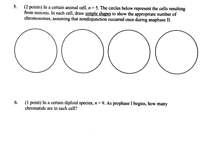
Solved In A Certain Animal Cell N 5 The Circles Below R
But the appearance of a tetrad changes in the different substages of prophase i read about these substages.
Prophase diagram simple. This is when the genetic fibers within the cells nucleus known as chromatin begin to condense and become tightly compacted together. Mitosis is the simplest of the two ways mitosis and meiosis in which the nucleus of a cell can divide as part of a process of whole cell division. The aster is an array of microtubules that radiates out from the centrosome towards the cell edge.
Mitosis begins at prophase with the thickening and coiling of the chromosomes. During interphase the parent cells chromosomes are replicated but they arent yet visible. The cell cycle phase which is the first stage of m phase of meiosis and mitosis and during which chromosomes condense and the two daughter centrioles and their asters migrate toward the poles of the cell.
Mitosis is the phase of the cell cycle where the nucleus of a cell is divided into two nuclei with an equal amount of genetic material in both the daughter nuclei. The nucleolus a rounded structure shrinks and disappears. During prophase they separate to provide microtubule centers in each.
In the upper diagram two tetrads are represented as two x shaped chromosomes associated side by side. Prophase is the first step of mitosis. It succeeds the g2 phase and is succeeded by cytoplasmic division after the separation of the nucleus.
Prophase is the first step of cell division in mitosis. The end of prophase is marked by the beginning of the organization of a group of fibres to form a spindle and the disintegration of the nuclear membrane. Prophase is the starting stage of cell division in eukaryotes.
Prophase in both mitosis and meiosis is recognized by the condensing of chromosomes and separation of the centrioles in the centrosomethis organelle controls the microtubules in the cell and each centriole is one half of the organelle. The four stages of mitosis prophase metaphase anaphase and telophase are shown and described below. Diagram also indicates the centromere region of a chromosome the narrow waist where the two sister chromatids are most tightly connected and the kinetochore a pad of proteins found at the centromere.
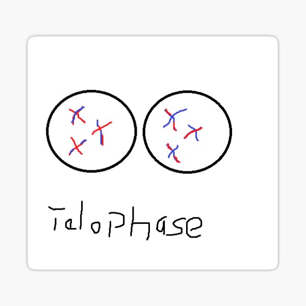
Prophase Stickers Redbubble

Mitosis Coloring Mitosis Cell Cycle Cell Cycle Activity

Learn For Free About Math Art Computer Programming Economics
Life Sciences Cyberbridge


Mitosis Article Cellular Division Khan Academy

The Cell Cycle Biology I
Early Prophase
Understanding The Process Of Cell Division Explained With Diagram

The 4 Mitosis Phases Prophase Metaphase Anaphase Telophase