Prophase Mitosis Microscope Slide
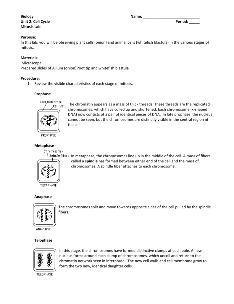
In the drawings below you can see the chromosomes in the nucleus going through the process called mitosis or division. During prophase the cytoskeleton composed of cytoplasmic microtubules begins to disassemble and the main component of the mitotic apparatus the mitotic spindle begins to form outside the nucleus at opposite ends of the cell.
Labeled Meiosis Microscope
Learn the microscope mitosis lab with free interactive flashcards.

Prophase mitosis microscope slide. Choose from 500 different sets of the microscope mitosis lab flashcards on quizlet. If you have a microscope 400x and a properly stained slide of the onion root tip or allium root tip you can see the phases in different cells. This video takes you through microscope images of cells going through mitosis and identifies the different phases under the microscope and on a micrograph.
If you have a microscope 400x and a properly stained slide of the onion root tip or allium root tip you can see the phases in different cells frozen in time. One of these processes involves the splitting of the chromosomes. In the drawings below you can see the chromosomes in the nucleus going through the process of mitosis or division.
The microscope and mitosis. Mitosis slide preparation from onion. The nuclear envelope breaks down and the nucleolus disappears.
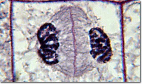
The first phase of mitosis is known as the prophase where the nuclear chromatin starts to become organized and condenses into thick strands that eventually become chromosomes. Microscope slides of mitosis cytokinesis. The cytoskeleton also disassembles and those microtubules form the spindle apparatus.
Prophase 1st stage of mitosis chromatin condenses down to form chromosomes the nuclear envelope breaks down and disappears centrosomes appear near the middle of the cell and move toward the poles of the cell. Prophase under a microscope during prophase the molecules of dna condense becoming shorter and thicker until they take on the traditional x shaped appearance.

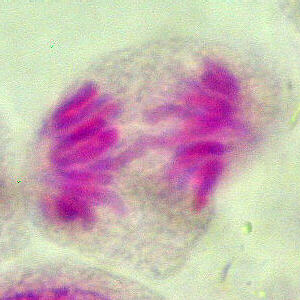
Prophase Whitefish Mitosis Whitefish Embryo Shows Chromosomes

Mitosis Lab William0912
Histology Laboratory Manual

Molecular Expressions Photo Gallery Mitosis

How To Identify Stages Of Mitosis Within A Cell Under A Microscope

Mitosis Lab Purpose

Mitosis Photographs
Anatomy A215 Virtual Microscopy

Fish Blastodisc Mitosis 400x Sec Petroarc International