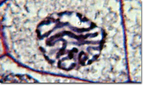
Prophase Plant Cell Under Microscope
As such it is a favorite in biology classrooms to show what a typical animal cell looks like. It is common to see photomicrographs of onion root cells when demonstrating how cell division takes place in plants.
Online Onion Root Tips

See a salamander grow from a single cell in this.

Prophase plant cell under microscope. In one particular cells nucleus the chromatin has condensed so much that it can be seen using a light microscope. The nucleoli primarily responsible for the production of ribosomal rna begin to disappear as the chromosomes condense. When observing the onion root tip cells for the stage of prophase the cells took on a brick like structure and within the cells small dots the nuclei can be seen.
Onions have larger chromosomes than most plants and stain dark. Mitosis is the mechanism that allows the nuclei of cells to split and provide each daughter cell with a complete set of chromosomes during cellular division. Fluorescence microscope image of two mouse cell nuclei in prophase scale bar is 5 µm.
Cell is a tiny structure and functional unit of a living organism containing various parts known as organelles. Top 10 most astonishing electron microscope pics in the. The apical meristem is an area of a plant where cell division takes place at a rapid rate.
The nuclear envelope breaks down and the nucleolus disappears. Learn the structure of animal cell and plant cell under light microscope. Identifying stages of mitosis under a microscope and on a.
Prophase is the first step of cell division in mitosis. The cells pictured below are located in the apical meristem of the onion root. The first stage of mitosis is known as prophase where the nuclear chromatin starts to become organized and condenses into thick strands that eventually become chromosomes observable in the optical microscope.
Prophase under a microscope during prophase the molecules of dna condense becoming shorter and thicker until they take on the traditional x shaped appearance. The chromosomes are easily observed through a compound light microscope. As it occurs after g2 of interphase dna has been already replicated when prophase begins.
See how a generalized structure of an animal cell and plant cell look with labeled diagrams. This video takes you through microscope images of cells going through mitosis and identifies the different phases under the microscope and on a micrograph. Cheek cells are easy to obtain and easy to see under a microscope.
This coupled with cytokinesis division of the cytoplasm occurs in all multicellular plants and animals to permit growth of the organism. The stage that the cell is currently in is prophase.
Mitosis Stages Of The Lily Microbehunter Microscopy


Mitosis In Onion Root Tips

Biology
Molecular Expressions Photo Gallery Mitosis
Uvqssxqb4jbrbm

Uvqssxqb4jbrbm


Lily Anther Late Prophase Of Meiosis C S 12 M Microscope Slide
Microscopy

The Cell 8 Cell Cycle Atlas Of Plant And Animal Histology