Prophase Cell Under Microscope
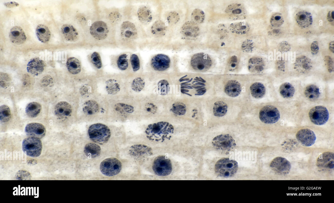
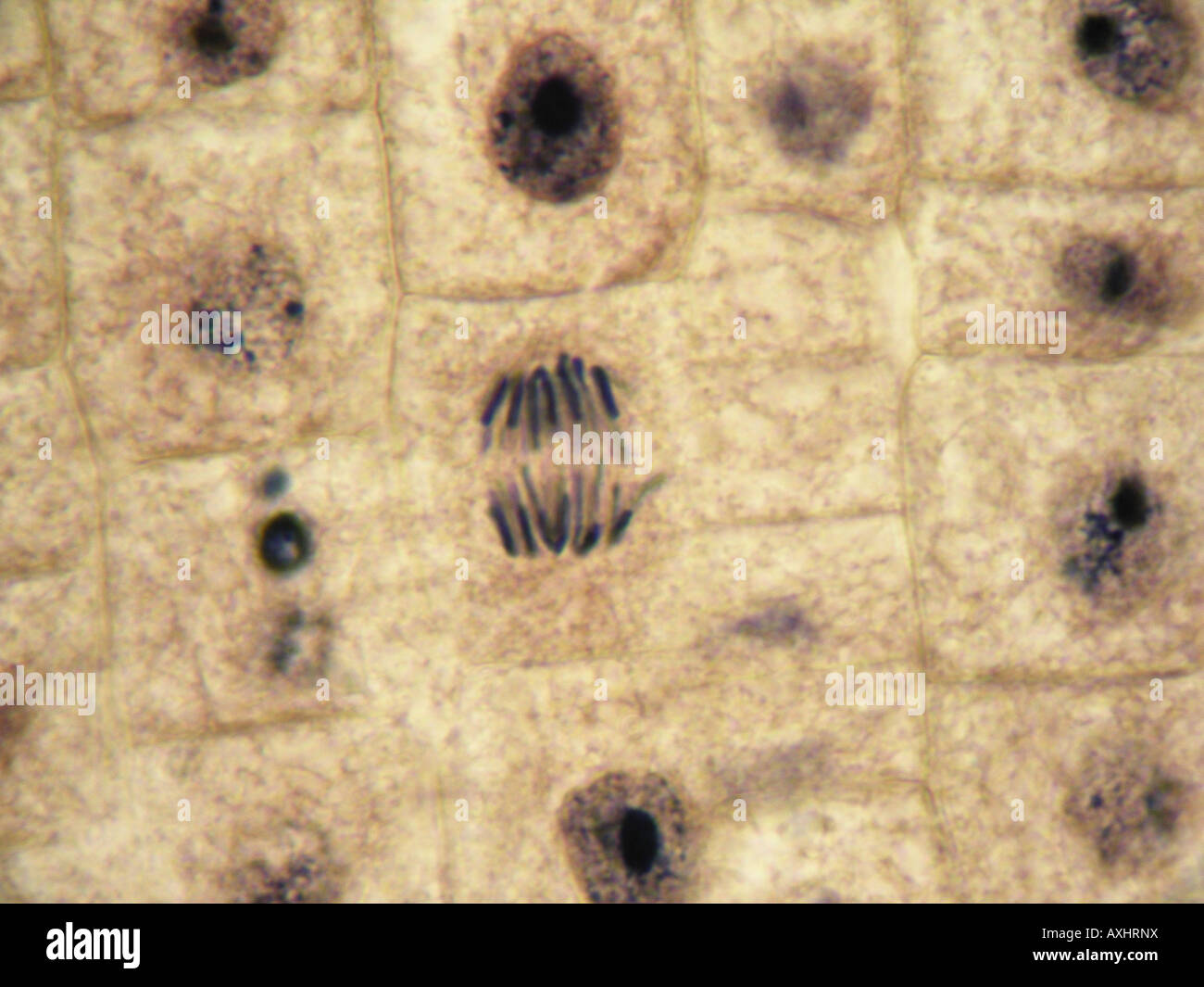
The nucleus will disappear freeing the chromosome pairs. Called metaphase the chromosomes line up in the center of the cell separate and become a pair of identical chromosomes.
Meiosis Anaphase Images Stock Photos Vectors Shutterstock
Is bright blue in a box shape.

Prophase cell under microscope. The cytoplasm starts separating cytokinesis. C large growths containing fluid. Chromosomes are lined up in the center of the cell.
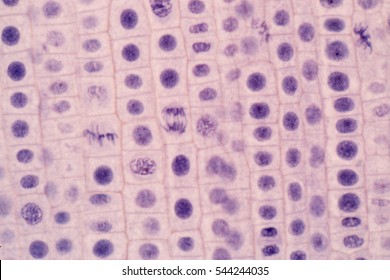
The invention of the microscope led to the identification of c cells are mostly water and therefore transparent so staining a molecule is said to be fluorescent if it absorbs light of o fix dehydrate section. During prophase the cytoskeleton composed of cytoplasmic microtubules begins to disassemble and the main component of the mitotic apparatus the mitotic spindle begins to form outside the nucleus at opposite ends of the cell. The onion root tip slide is included free in your slide kit when you purchase a microscope from microscope world.
During prophase the nuclear envelope starts to break down and all the chromosomes start to coil up in the center of the cell. Prophase under a microscope during prophase the molecules of dna condense becoming shorter and thicker until they take on the traditional x shaped appearance. At the start of prophase the chromosomes condense and can now be seen under a microscope.
The nucleoli primarily responsible for the production of ribosomal rna begin to disappear as the chromosomes condense. 3 metaphase is the middle stage at which point all the chromosome pairs line up in the center of the cell along spindle fibers that pull to either side of the cell. The cytoskeleton also disassembles and those microtubules form the spindle apparatus.
The first stage of mitosis is known as prophase where the nuclear chromatin starts to become organized and condenses into thick strands that eventually become chromosomes observable in the optical microscope. The nuclear envelope breaks down and the nucleolus disappears. The photomicrograph below depicts the initial chromosome condensation at the beginning of prophase early prophase when the nucleolus is still intact.
The centrioles begin to migrate toward the far ends of the cell while the mitotic spindle forms. Cheek cells are easy to obtain and easy to see under a microscope. Is a brown box with darker brown spots.
Called prophase the dna molecules of the chromosomes condense. B cells that are much smaller than normal cells. As such it is a favorite in biology classrooms to show what a typical animal cell looks like.
The pairs of chromatids that make up the chromosomes separate from each other and are pulled to two opposite ends of the cell. The phases of mitosis. A scientist who observes cancer cells under a microscope would likely see a orderly rows of cells.
Nuclear membranes form around both sets of chromosomes and the chromosomes uncoil.
Microscopy

Mitosis Review Looking At The Pictures Below And To The Right Please


The Miracle Of Mitosis Cell Division Microscope It Out

1 6 Skill Identifying Stages Of Mitosis Under A Microscope And On
Mitosis Cell Division In Onion Root Tip Brightfield

Microscopic Images Of Chromosomes At Different Stages Of Cell
Root Tip Of Onion Show Mitosis Cell In The Root Tip Under The
Mitosis Anaphase In Onion Tissue At 1000x Under Optical
Https Www Sps186 Org Downloads Basic 461381 Plant 20cell 20mitosis Pdf