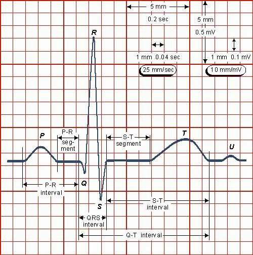
P Q R S T Wave
Large waves are referred to by their capital letters q r s and small waves are referred to by their lower case letters q r s. A q wave is any downward deflection immediately following the p wave.

Pqrst Wave Nursing Mnemonics Nurse Nursing Students
Arrhythmias the normal ekg consists of repetitive series of p q r s and t waves which conform to established standards for size and shape and occur 60 100 times each minute.

P q r s t wave. This spike is called the qrs complex. Any negative wave occurring after a positive wave is an s wave. The p wave represents atrial depolarization depolarization is a big fancy word for contraction.
The t wave follows the s wave and in some cases an additional u wave follows the t wave. The ecg learning center explains that the p wave represents the depolarization of the right and left atria. The p wave qrs complex and the t wave represent electrical activity in the heart on an electrocardiogram.
The first little hump or bump you see is known as the p wave. Remember from the electrical conduction lecture that the sa node is responsible for this. The next area you see is a big spike.
An r wave follows as an upward deflection and the s wave is any downward deflection after the r wave. If these conditions prevail the heart is in normal sinus rhythm. Figure 5 shows examples of the naming of the qrs complex.

Morphology Of One Pqrst Complex Of The Ecg With Big T Wave

Image Result For Pqrst Wave Qrs Complex Nursing Fun Nursing Study

Qrs Complex Wikipedia

Detection Of P Q R S T Waves A Sample Of Ecg Signal A Original

The Normal Ecg The Student Physiologist

Ecg Traces Flashcards Quizlet

Electrical Signals Of The Heart

Ekg Waves Complexes Straight Lines And Intervals Labeling And
1 The Pqrst Wave Structure Of An Ecg Segment Download